Cardiac T2*
Cardiac T2* for Iron Assessment
The Liver and Heart in Iron Overload
Changes in liver iron concentration (LIC) generally precede changes in cardiac iron loading. As cardiac iron loading increases so does the risk of iron-induced arrhythmia and cardiac failure. Cardiac iron-loading can be assessed by MRI measurement of cardiac T2* (cardiac T2* is globally recognised as the gold standard in the assessment of cardiac iron load via T2* measurement). Iron-induced damage to heart tissue may be mitigated if appropriate and timely chelation therapy is commenced.
Every site globally that is set up for Resonance Health Cardiac T2* acquires images in a standardised manner, all analysis is performed in a stringently quality controlled core laboratory, and results are directly comparable from site to site and across time.
Regular assessment of body iron overload through the FerriScan and Cardiac T2* Dual Analysis Service enables better informed decisions on the management of patients at risk of iron-induced organ damage.
The Cardiac T2* Report
Cardiac T2* for Iron Assessment is a service that is offered separately or as part of a dual analysis package with FerriScan to provide more comprehensive information regarding body iron stores.
Cardiac T2* through Resonance Health, has the following features:
- Provides important information for assessing the risk of cardiac failure or arrhythmia
- Cardiac T2* is the most widely accepted MRI-based method for assessing heart iron loading
- Cardiac T2* is charged per scan only and can be established on most 1.5T MRI scanners
Why use MRI and Cardiac T2*
MRI is an ideal method for acquiring data for the assessment of iron overload. Iron produces disturbances in the magnetic field and the greater the iron load, the greater the signal decay rate. The figure below shows a short axis mid-ventricular T2* image of the heart. Echo times from <3ms to >16ms are used to assess the signal intensities in the septum from which the cardiac T2* (R2*) can be calculated.

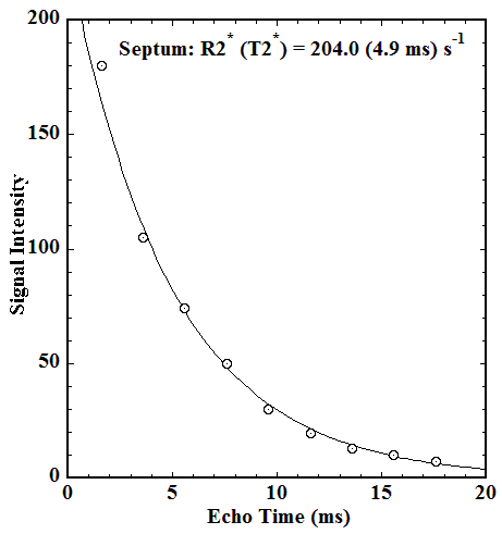
Figure. Note that the septum outline is for illustrative purposes only. Resonance Health endeavors to maximise the septum region. However, the area selected for analysis will vary depending on the level of artifact.
There are several published methods for acquiring data for cardiac T2* analysis which may vary across MRI scanners. These techniques generally involve a breath-hold (usually 8-12 seconds) multi echo gradient echo pulse sequence to acquire a series of images at different echo times with either dark blood or bright blood method. An ECG-gated sequence is used to compensate for cardiac motion.
The cardiac T2* imaging protocol provided by your MRI scanner manufacturer should be used with modification defined by Resonance Health’s Cardiac T2* service.
It is recommended to use Cardiac T2* through Resonance Health to enable a patients’ results to be compared over time.
Cardiac T2* Resources
Click the link(s) below to access our resources. Need something else? Please contact our team at [email protected] for further information.